Overview and Epidemiology
Babesiosis in cattle is a tick-borne disease caused primarily by Babesia bovis and Babesia bigemina. It’s transmitted via bites from ixodid ticks (e.g., Rhipicephalus spp.) and, less commonly, iatrogenically (e.g., contaminated needles or transfusions) (cfsph.iastate.edu).
This disease, also known as Texas cattle fever or redwater, is especially severe in non-immune adult cattle with high fever, hemolytic anemia, hemoglobinuria, jaundice, and potentially cerebral signs (“cerebral babesiosis”) (en.wikipedia.org).
Babesiosis – Summary
Canine Babesiosis
Clinical Signs:
- Vary from mild to severe
- Fever, lethargy, anorexia
- Pale or icteric mucous membranes
- Splenomegaly, ± lymphadenopathy
Etiologic Agents:
- Babesia canis vogeli (large form) – seen in Greyhounds
- Babesia gibsoni (small form) – common in Pit Bulls, Tosa Inus
Transmission:
- Ticks (Rhipicephalus, Dermacentor spp.)
- Blood transfusion
- Dog fights, contaminated instruments, vertical transmission
Diagnosis:
- Giemsa-stained smear: organisms in RBCs
- CBC: thrombocytopenia, ± regenerative anemia
- Chemistry: hyperglobulinemia, ± bilirubinemia
- Serology (IFA ≥1:64), PCR, positive Coombs’ test
- Radiography: splenomegaly
Treatment:
- B. canis vogeli: Imidocarb (pretreat with atropine)
- B. gibsoni: Atovaquone + Azithromycin
- Supportive care: blood transfusion, IV fluids
Bovine Babesiosis
Clinical Signs:
- Fever, anorexia, pale mucous membranes
- Weakness, reluctance to move
- ± Icterus, abortion, neurologic signs (B. bovis only)
Etiologic Agents:
- Babesia bigemina and Babesia bovis
- Transmitted by Rhipicephalus spp. ticks
Diagnosis:
- Giemsa-stained blood smear: intraerythrocytic parasites
- CBC: anemia, hemoglobinemia (esp. B. bigemina)
- Serology (IFA, ELISA)
- Necropsy: enlarged spleen, dark kidneys
Treatment:
- Imidocarb or Diminazene
- Blood transfusion, IV fluids, NSAIDs
Equine Babesiosis (Equine Piroplasmosis)
Clinical Signs:
- Fever, weakness, anorexia
- Icterus, petechiae, colic, sudden death
- Exercise intolerance
Etiologic Agents:
- Babesia caballi, Theileria equi
- Spread by ticks and iatrogenic blood transfer
Diagnosis:
- Blood smear, PCR, serology
Treatment:
- Imidocarb (difficult to eliminate carrier state)
- Blood transfusion, oxygen therapy
Key Points
- Dogs: Not zoonotic; co-infections with other tick-borne pathogens possible
- Bovine: Severe signs with B. bovis; hemoglobinuria with B. bigemina
- Equine: REPORTABLE disease in the U.S.; foreign animal disease
- Human: Babesia divergens serious in splenectomized individuals
Clinical Signs
- Acute high fever (≥ 106 °F), depression, inappetence, pale mucous membranes
- Icterus, hemoglobinuria (“red-water”), tachypnea, tachycardia
- Nervous signs in B. bovis (ataxia, incoordination, seizures)
- Abortion in pregnant cows and temporary infertility in bulls (researchgate.net, en.wikipedia.org, sciencedirect.com)
Diagnostic Modalities
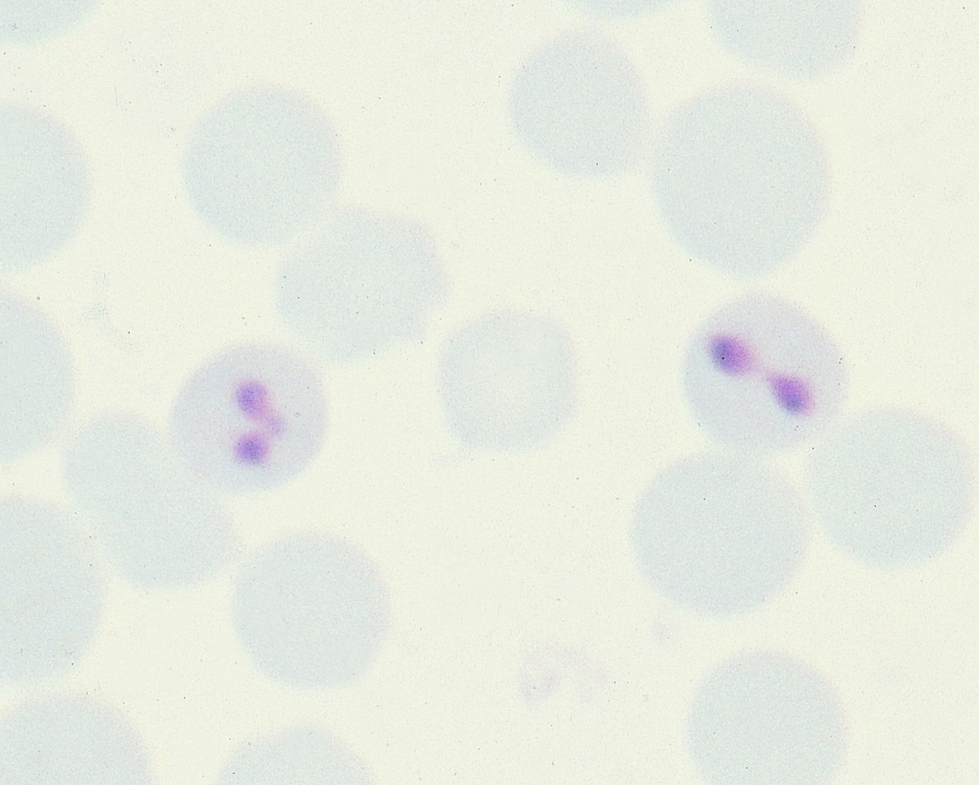
- Blood smear microscopy: Identification of pyriform or paired merozoites in RBCs using Giemsa/Wright–Giemsa stain. B. bovis are small, obtusely angled pairs; B. bigemina are larger, acutely angled (merckvetmanual.com).
- PCR: High sensitivity for species detection, including in carrier animals (pmc.ncbi.nlm.nih.gov).
- Serology (IFAT, ELISA): Identifies exposure; less useful in acute diagnosis (en.wikipedia.org).
- Laboratory findings: Hemolytic anemia, hemoglobinemia, elevated bilirubin; possible DIC in severe cases (en.wikipedia.org).
Interpretation of the Image Carousel
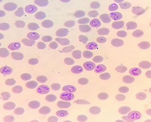
- Image 1 (turn0image0) shows pyriform merozoites inside bovine RBCs.
- Image 2 (turn0image1) demonstrates paired B. bovis organisms at obtuse angles.
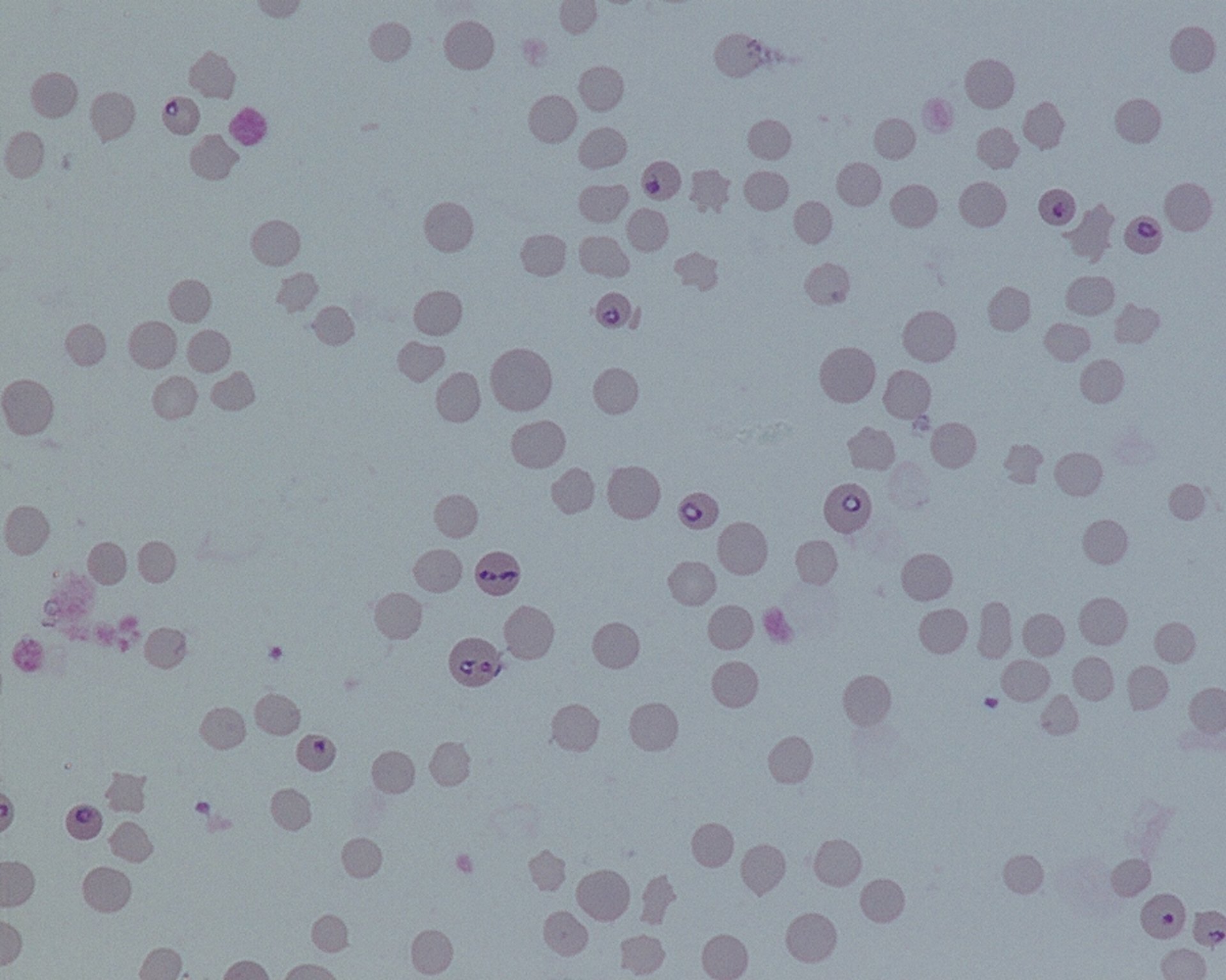
- Image 3 (turn0image2) highlights small merozoites with red arrows pointing to ring forms.

- Image 4 (turn0image3) presents two infected RBCs typical of Babesia bovis infection.
Treatment
- Imidocarb dipropionate or diminazene aceturate are the main anti-protozoal treatments (en.wikipedia.org, en.wikipedia.org, cell.com, merckvetmanual.com).
- Supportive therapy with NSAIDs, IV fluids, and blood transfusions as needed (merckvetmanual.com).
Prevention and Control
- Tick control: Use of acaricides, pasture management, and integrated pest strategies (merckvetmanual.com).
- Vaccination: Live-attenuated vaccines exist in endemic areas (not available in the U.S.) (en.wikipedia.org).
- Biosecurity: Prevent transfusion- or equipment-mediated transmission.
- Carrier detection: Use PCR or serology to manage carriers and reduce transmission (en.wikipedia.org, merckvetmanual.com).
NAVLE‑Style Multiple‑Choice Questions
Q1: Diagnosis
A 4-year-old beef cow from a subtropical region exhibits fever (105 °F), hemoglobinuria, icterus, and neurological signs. Blood smear under oil immersion shows small pyriform merozoites within RBCs arranged as obtuse‑angled pairs. What is the most likely diagnosis?
A. Anaplasmosis
B. Babesiosis
C. Theileriosis
D. Leptospirosis
E. Hemolytic anemia from immune-mediated disease
Correct Answer: B. Babesiosis
Explanation: Fever, hemoglobinuria, and intracellular pyriform merozoites are consistent with Babesia spp., particularly B. bovis (merckvetmanual.com, my.clevelandclinic.org).
Q2: Treatment
A cow confirmed with acute bovine babesiosis has severe anemia and hemoglobinuria. What is the most appropriate treatment protocol?
A. Oxytetracycline + blood transfusion
B. Imidocarb dipropionate + supportive care
C. Long‑acting penicillin only
D. Doxycycline + NSAIDs
E. Ivermectin and vitamin supplementation
Correct Answer: B. Imidocarb dipropionate + supportive care
Explanation: Imidocarb or diminazene are effective treatments. Supportive therapy is essential in cases of hemolytic anemia and hemoglobinuria (en.wikipedia.org, merckvetmanual.com).
Q3: Prevention/Transmission
A herd of cattle develops babesiosis after the introduction of new animals. Aside from vaccination, which control measure would be most effective?
A. Frequent use of broad‑spectrum antibiotics
B. Isolation of new arrivals and tick control programs
C. High‑fiber dietary supplementation
D. Administering live A. centrale vaccine
E. Routine deworming
Correct Answer: B. Isolation of new arrivals and tick control programs
Explanation: Preventing disease spread requires quarantine, acaricidal tick control, and proper management .
References
- Merck Veterinary Manual – Babesiosis in Animals (merckvetmanual.com)
- MSD Veterinary Manual – Babesia bovis, blood smear image (msdvetmanual.com)
- CDC Diagnostic Bench Aids – Babesia morphology (cdc.gov)
- CFSPH Iowa State – Bovine babesiosis outbreak control (cfsph.iastate.edu)
- Texas A&M, ScienceDirect, and Cell Press – Parasite biology and treatment protocols (cell.com)