Overview
Calf enteritis, commonly referred to as calf scours, is a leading cause of morbidity and mortality in neonatal calves, particularly within the first month of life. The condition is characterized by diarrhea resulting from various infectious agents, leading to dehydration, electrolyte imbalances, and, if untreated, death. Prompt recognition and management are crucial for calf survival.Merck Veterinary Manual
Etiology
Infectious Agents:
- Bacterial:
- Enterotoxigenic Escherichia coli (ETEC): Primarily affects calves in the first week of life. ETEC possesses fimbrial antigens (e.g., F5/K99) facilitating adherence to intestinal villi, leading to secretory diarrhea.
- Salmonella spp.: Typically affects calves aged 2–12 weeks. It causes enteritis and can lead to systemic infection.
- Clostridium perfringens: Produces toxins causing hemorrhagic enteritis, often leading to sudden death. Merck Veterinary Manual
- Viral:
- Rotavirus: Common in calves aged 5–14 days. It damages the villous tips of enterocytes, leading to malabsorption.
- Coronavirus: Affects both small and large intestines, leading to villous atrophy and diarrhea. DigitalCommons+4PMC+4vetsci.org+4Merck Veterinary Manual
- Protozoal:
- Cryptosporidium parvum: Typically affects calves aged 5–35 days. It attaches to the intestinal epithelium, causing villous atrophy and malabsorption. Merck Veterinary Manual
Non-Infectious Factors:
- Inadequate colostrum intake leading to failure of passive transfer.
- Poor hygiene and environmental conditions.
- Nutritional factors, such as overfeeding or abrupt dietary changes.
Clinical Signs
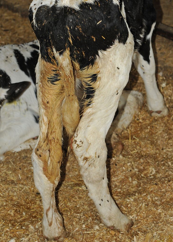
- Watery to pasty diarrhea, possibly containing blood or mucus.
- Dehydration indicators: sunken eyes, skin tenting, dry mucous membranes.
- Weakness, depression, and reluctance to stand or nurse.
- Hypothermia or fever, depending on the causative agent.
- In severe cases, recumbency and death.
Classic Case
- Age: 2–10 days old
- Signs:
- Diarrhea
- Lethargy
- Depression
- Hypothermia
- Signs of sepsis
Diagnosis
Colostrum Assessment
- Check for adequate passive transfer
- Use refractometer:
- Total protein >5.5 g/dL indicates sufficient colostrum intake
- Use refractometer:
Laboratory Diagnostics
- Fluid and acid-base status:
- Assess blood pH, bicarbonate, base excess, and glucose to guide fluid therapy in severely dehydrated calves
Pathogen Identification
- Salmonella spp.:
- Culture feces at least 5 times for confirmation
- Giardia spp.:
- Fecal flotation and direct smear
- Cryptosporidium spp.:
- Acid-fast stain or immunoassay on fecal samples
- Bovine Viral Diarrhea (BVD):
- PCR on ear notch tissue or blood
- Rotavirus:
- Fecal Rotazyme test
- Coronavirus:
- Fluorescent antibody test on duodenal/jejunal tissue
Treatment
Fluid Therapy
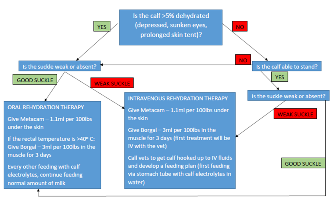
- If ≥5–7% dehydrated and severely ill:
- Use intravenous fluids (IVF)
- Select fluid type based on acid-base status (commonly sodium bicarbonate + dextrose)
- If <5–7% dehydrated:
- Use enteral fluids:
- Electrolyte replacers
- Continue milk feeding
- Use enteral fluids:
Additional Therapies
- Antimicrobials: As indicated (especially if sepsis suspected)
- Anti-inflammatories: Supportive care
Prevention
- Calf Management:
- Keep calving areas and hutches clean
- Provide high-quality colostrum:
- 1 gallon per 100 lb calf at the first feeding
Key Points / Pearls
- Multifactorial outbreaks:
- Most diarrhea outbreaks involve multiple pathogens
- Colostrum is critical to prevent early diarrhea and sepsis
- Monitor for complications:
- Secondary conditions like Lumpy Jaw or Wooden Tongue may appear in poorly managed or immune-compromised calves
NAVLE-Style Multiple-Choice Questions
Question 1: Etiology
Clinical Scenario:
A 5-day-old calf presents with profuse watery diarrhea. Fecal antigen testing is positive for rotavirus.
Question:
Which of the following best describes the pathogenesis of rotavirus in calves?
A. Invasion of the intestinal mucosa leading to hemorrhagic enteritis.
B. Attachment to enterocytes causing secretory diarrhea via enterotoxin production.
C. Destruction of villous enterocytes leading to malabsorption.
D. Colonization of the colon resulting in pseudomembranous colitis.
E. Systemic bacteremia leading to endotoxemia.Merck Veterinary Manual
Correct Answer: C. Destruction of villous enterocytes leading to malabsorption.
Explanation: Rotavirus infects and destroys the mature enterocytes lining the villi of the small intestine, leading to villous atrophy and malabsorption, resulting in diarrhea. Merck Veterinary Manual
Question 2: Diagnosis
Clinical Scenario:
A 10-day-old calf exhibits diarrhea, depression, and dehydration. Fecal culture identifies Salmonella Typhimurium.
Question:
Which of the following additional clinical signs is most consistent with Salmonella infection in calves?
A. Neurological signs such as seizures.
B. Hemorrhagic diarrhea with mucosal shreds.
C. Chronic weight loss without diarrhea.
D. Respiratory distress due to pneumonia.
E. Skin lesions and alopecia.
Correct Answer: B. Hemorrhagic diarrhea with mucosal shreds.
Explanation: Salmonella infections in calves often cause severe enteritis, leading to bloody diarrhea with mucosal shreds due to intestinal mucosal damage. Merck Veterinary Manual
Question 3: Treatment
Clinical Scenario:
A 7-day-old calf with diarrhea is 8% dehydrated but still able to stand and has a suckle reflex.
Question:
What is the most appropriate initial treatment for this calf?
A. Immediate intravenous fluid therapy.
B. Oral rehydration therapy with electrolyte solutions.
C. Administration of broad-spectrum antibiotics only.
D. Withholding milk feeding for 48 hours.
E. Administration of anti-diarrheal medications to reduce motility.Details are here
Correct Answer: B. Oral rehydration therapy with electrolyte solutions.
Explanation: In calves with moderate dehydration that are still standing and have a suckle reflex, oral rehydration therapy is effective and less invasive. Intravenous fluids are reserved for more severe cases. PMC
Visual References
To enhance understanding, here are some visual references related to calf enteritis:
- Clinical Signs of Calf Scours: Image depicting sunken eyes and dehydration signsPro Earth Animal Health
- Dehydration Assessment Chart: Diagram illustrating dehydration levels and corresponding clinical signstommythevet.ie
- Pathogen Identification Table: Chart categorizing pathogens by age of onset and clinical featuresScribd+3Bovine Veterinarian+3Bovine Veterinarian+3
References
- Merck Veterinary Manual: [Diarrhea in Neonatal Ruminants](https://www.merckvetmanual.com/digestive-system/intestinal-diseases-in