Overview
Cecal disorders in cattle primarily involve cecal dilatation, cecal volvulus (torsion), and cecal retroflexion. These conditions can lead to partial or complete intestinal obstruction, resulting in significant clinical signs and requiring prompt diagnosis and management.
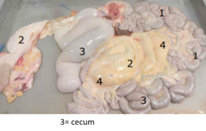
Cecum here is 3 but you should know the anatomy of all of the followings… large intestine surgeries
Etiology
- Cecal Dilatation: Often associated with high-concentrate, low-fiber diets leading to decreased motility and gas accumulation. Hypocalcemia and other metabolic disturbances may also contribute.Merck Veterinary Manual
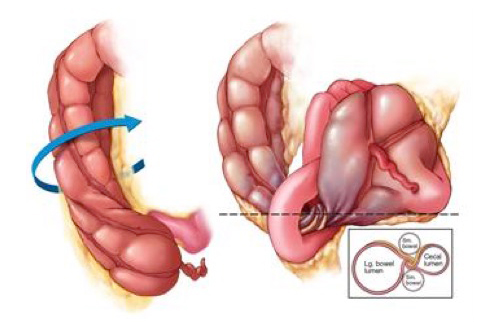
- Cecal Volvulus (Torsion): Occurs when the cecum rotates around its longitudinal axis, potentially compromising blood flow and leading to ischemia (more details)
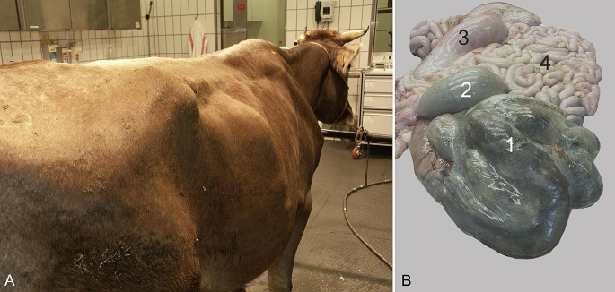
A large bulge in the right flank (A) and postmortem findings (B) in a 4-year-old Brown Swiss cow with torsion of the spiral colon (also see video). The gyri of the spiral colon (1) and the proximal loop of the ascending colon (2) are congested and severely dilated and have a dark bluish-green discolouration. The caecum (3) is dilated but otherwise normal. The small intestines are mildly to moderately congested but otherwise normal. Reference
- Cecal Retroflexion: The cecum folds dorsally or ventrally in the ileocecal region, causing a cranial orientation of the apex and possible obstruction. Learn more at BioMed Central and BMC Central.
Clinical Signs
- Decreased or absent fecal output.Merck Veterinary Manual
- Anorexia and decreased milk production.Merck Veterinary Manual+2Merck Veterinary Manual+2Wikipedia+2
- Abdominal distension, particularly in the right flank.
- Signs of colic: restlessness, kicking at the abdomen, treading, and teeth grinding. (Merck Veterinary Manual)
- On rectal examination: distended cecum palpable in the right pelvic area; in cases of torsion or retroflexion, the cecum may be displaced cranially.BioMed Central
Diagnosis
- Rectal Examination: Primary diagnostic tool; allows palpation of the distended cecum in approximately 95% of cases. BioMed Central
- Ultrasonography: Useful when rectal palpation is inconclusive; can identify gas-distended cecum and assess for torsion.
- Laboratory Tests: May reveal electrolyte imbalances (e.g., hypocalcemia), dehydration, and metabolic alkalosis.
Treatment
- Medical Management (for uncomplicated cecal dilatation):
- Administration of parasympathomimetic agents (e.g., neostigmine) to stimulate motility.More details at BioMed Central
- Correction of electrolyte imbalances and dehydration.
- Monitoring for progression or resolution.
- Surgical Intervention (indicated for torsion, retroflexion, or non-responsive dilatation):
- Right flank laparotomy to decompress the cecum.
- Typhlotomy (surgical opening of the cecum) to evacuate contents.(check more here)
- Cecal amputation may be necessary in cases of necrosis or recurrent dilatation.BioMed Central
Prognosis
- Favorable in cases of simple dilatation with prompt medical management.
- Guarded to poor in cases involving torsion or necrosis, even with surgical intervention.
NAVLE-Style Multiple-Choice Questions
Question 1:
A 5-year-old Holstein cow presents with decreased appetite, reduced milk production, and scant fecal output. Rectal examination reveals a gas-distended viscus in the right pelvic area. What is the most likely diagnosis?
A. Left displaced abomasum
B. Cecal dilatation
C. Intussusception
D. Right displaced abomasum
E. Rumen tympany
Correct Answer: B. Cecal dilatation
Explanation: The presence of a gas-distended viscus in the right pelvic area on rectal examination is characteristic of cecal dilatation.
Question 2:
Which of the following is the most appropriate initial treatment for uncomplicated cecal dilatation in cattle?
A. Immediate surgical intervention
B. Administration of neostigmine and correction of electrolyte imbalances
C. Withholding feed and water for 48 hours
D. Administration of broad-spectrum antibiotics
E. Rumenotomy
Correct Answer: B. Administration of neostigmine and correction of electrolyte imbalances
Explanation: Medical management with parasympathomimetic agents like neostigmine, along with correcting electrolyte imbalances, is effective for uncomplicated cecal dilatation.BioMed Central
Question 3:
In cases of cecal volvulus, which of the following clinical signs is most likely to be observed?
A. Profuse watery diarrhea
B. Bradycardia
C. Severe abdominal pain with signs of colic
D. Hypersalivation
E. Left-sided abdominal distension
Correct Answer: C. Severe abdominal pain with signs of colic
Explanation: Cecal volvulus often presents with acute, severe abdominal pain and signs of colic due to compromised blood flow and intestinal obstruction (learn more)
References
- BMC Veterinary Research. (2012). Clinical findings and treatment in cattle with caecal dilatationBioMed Central
- Merck Veterinary Manual. (2021). Acute Intestinal Obstructions in Large AnimalsMerck Veterinary Manual
- University of Minnesota. (n.d.). Cecal dilation – Large Animal Surgery – Supplemental Notesopen.lib.umn.edu+3open.lib.umn.edu+3open.lib.umn.edu+3
- Veterinary World. (2018). Differential diagnosis and surgical management of cecal dilatationVeterinary World
- Journal of the American Veterinary Medical Association. (1986). Cecal dilatation and volvulus in dairy cows: 84 cases (1977–1983)AVMA Journals