Overview
Histopathologic findings of intestine and lymph nodes with with Johne’s disease. (A) Accumulation of inflammatory cells in the lamina propria of the small intestine. (B) Higher magnification of (A) showing large macrophages (arrowheads) and lymphocytes (arrows) accumulated in the lamina propria. (C) Mesenteric lymph node. Note the multinucleated giant cell (arrow). A-C, H-E staining. A and C: Scale bars = 100 µm, B: Scale bar = 50 µm.
Paratuberculosis, also known as Johne’s disease, is a chronic, contagious granulomatous enteritis caused by Mycobacterium avium subsp. paratuberculosis (MAP). Infection occurs early in life—usually within a few months of birth—but clinical signs often appear only after several years. It’s characterized by weight loss, diarrhea, and eventual euthanasia due to poor prognosis. (merckvetmanual.com)
Etiology & Pathogenesis
- Agent: MAP, a hardy acid-fast bacillus shed in feces, colostrum, and milk. (merckvetmanual.com)
- Transmission: Primarily fecal-oral; calves are at greatest risk soon after birth. The organism targets Peyer’s patches in the lower ileum, multiplying within macrophages and causing chronic granulomatous inflammation. (merckvetmanual.com)
Clinical Signs
- Subclinical phase: Asymptomatic but shedding bacteria.
- Clinical phase: Usually in mature cattle (2–6 years): progressive weight loss, intermittent to chronic watery diarrhea (non-bloody), rough hair coat, bottle jaw (submandibular edema), reduced milk yield. (en.wikipedia.org)
Pathology
- Marked thickening and corrugation of ileal/colonic mucosa; enlarged mesenteric lymph nodes. (pmc.ncbi.nlm.nih.gov)
- Microscopically: granulomatous enteritis with epithelioid macrophages and multinucleated giant cells infiltrating mucosa/submucosa. Acid-fast MAP within macrophages. (merckvetmanual.com)
Diagnosis
- Ante-mortem:
- Fecal PCR or culture (though intermittent shedding reduces sensitivity).
- Serum ELISA for herd-level screening.
- Acid-fast staining of intestinal biopsies. (merckvetmanual.com)
- Post-mortem:
- Gross and histopathologic confirmation: intestinal thickening, lymphadenopathy, granulomatous lesions with acid-fast organisms.
Treatment & Control
- Treatment: Not practical—no antibiotic cures; supportive care only.
- Control measures:
- Birth in clean environments, early removal from dam, feeding colostrum from test-negative cows.
- Maintain separate housing for young calves until >1 year.
- Test-and-cull programs with yearly herd testing. (merckvetmanual.com)
Zoonotic Consideration
- MAP has been detected in humans with Crohn’s disease, but evidence of zoonosis remains inconclusive. Pasteurizing milk remains a practical mitigation step. (merckvetmanual.com)
NAVLE-Style Multiple-Choice Questions
Question 1: Diagnosis
A 4-year-old dairy cow presents with chronic watery diarrhea, weight loss, bottle jaw, and reduced milk production. Fecal PCR is positive for MAP. What is the most likely histopathologic finding in the ileum?
A. Villous atrophy with crypt hyperplasia
B. Granulomatous inflammation with acid-fast bacilli within macrophages
C. Erosions with neutrophilic exudate and fibrin
D. Hypertrophy of Brunner’s glands
E. Lymphocytic cholangiohepatitis
Correct Answer: B
Explanation: Johne’s disease causes granulomatous enteritis; epithelioid macrophages with acid-fast MAP confirm diagnosis. (askjpc.org)
Question 2: Treatment & Control
Which of the following management practices is most effective in controlling Johne’s disease in a dairy herd?
A. Feeding low-protein diets to adult cattle
B. Removing calves at birth and feeding colostrum from test-negative dams
C. Mass vaccination using live MAP vaccine
D. Administering ivermectin to all heifers at weaning
E. Housing cows on pasture year-round
Correct Answer: B
Explanation: Calves born to infected dams ingesting contaminated milk are at high risk. Removing them and feeding colostrum from test-negative cows reduces transmission significantly. (aphis.usda.gov, merckvetmanual.com)
Question 3: Epidemiology
What is a characteristic feature of MAP infection at the herd level?
A. Acute hemorrhagic enteritis in all age groups
B. High morbidity in calves under 6 months old with severe symptoms
C. Young infection with adult-onset clinical signs
D. Zoonotic spread to farmworkers is inevitable
E. Effective antibiotic treatment is available
Correct Answer: C
Explanation: Cattle are infected as calves, but symptoms generally manifest years later. Morbidity and mortality are low; infection is chronic. (pmc.ncbi.nlm.nih.gov, askjpc.org)
Classic Case
- Most infected cattle are subclinical for years
- Clinical disease often presents between ages 3–5 years
Stages of Disease:
- Stage 1:
- Young animals
- Infected but asymptomatic
- Not shedding
- Negative on diagnostic tests
- Stage 2:
- Older animals
- Still asymptomatic
- Shed organism in feces
- Positive on culture and ELISA
- Stage 3:
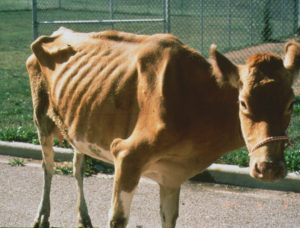
- Thin, adult cow (3–5 years)
- Voluminous, watery diarrhea
- Decreased milk production
- Brisket edema, enlarged mesenteric lymph nodes
- No neurologic deficits (e.g., no proprioceptive loss, facial paralysis, or Horner’s syndrome)
Diagnosis
- Individual animal:
- Fecal culture: gold standard
- PCR, rectal mucosa histopathology also useful
- Herd-level:
- Pooled fecal cultures
- Pooled PCR for early detection
- Herd surveillance:
- Serum or milk ELISA
- Serum more sensitive; both highly specific
- Serum or milk ELISA
Treatment
- No effective treatment
- Affected animals should be euthanized and reported
Control & Prevention
- Separate calves from manure and use clean feeding equipment
- Do not pool colostrum
- Conduct annual ELISAs
- Maintain a closed or clean young replacement herd
Key Points
- Can cause early-term abortion
- For each clinical cow (Stage 3):
- Expect 3–4 animals in Stage 2
- And 10–15 animals in Stage 1
- Often co-infects with enteric pathogens, such as E. coli